CASE20250513_001
A Rare Case of Residual Atrial Septal Defect Following Device Closure
By Chanikarn Kanaderm
Presenter
Chanikarn Kanaderm
Authors
Chanikarn Kanaderm1
Affiliation
Central Chest Institute of Thailand, Thailand1,
View Study Report
CASE20250513_001
Atrial Septal Defect (ASD) Closure - Atrial Septal Defect (ASD) Closure
A Rare Case of Residual Atrial Septal Defect Following Device Closure
Chanikarn Kanaderm1
Central Chest Institute of Thailand, Thailand1,
Clinical Information
Relevant Clinical History and Physical Exam
A 38-year-old woman underwent ASD device closure 4 years ago. Early post-op TTE at 6 and 12 months showed no residual shunt. She was lost to follow-up for 3 years due to COVID-19 endermic. She later developed palpitations and shortness of breath on exertion. On physical exam, a grade II/VI systolic murmur was noted at the left sternal border, second intercostal space.
Relevant Test Results Prior to Catheterization
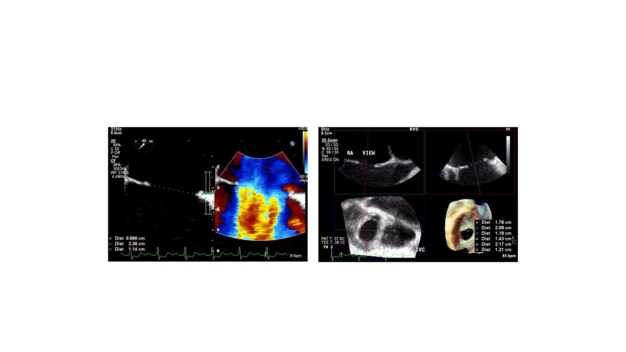
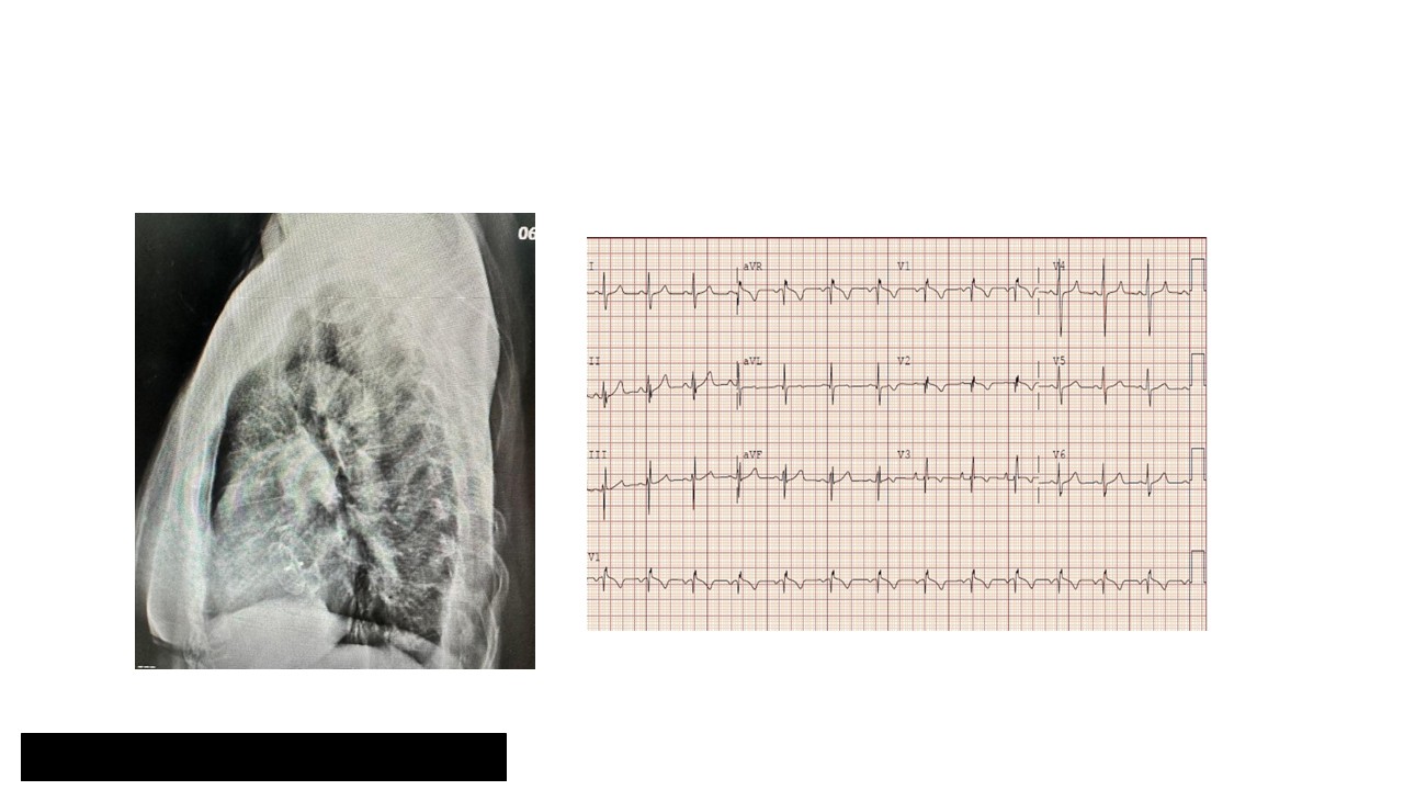
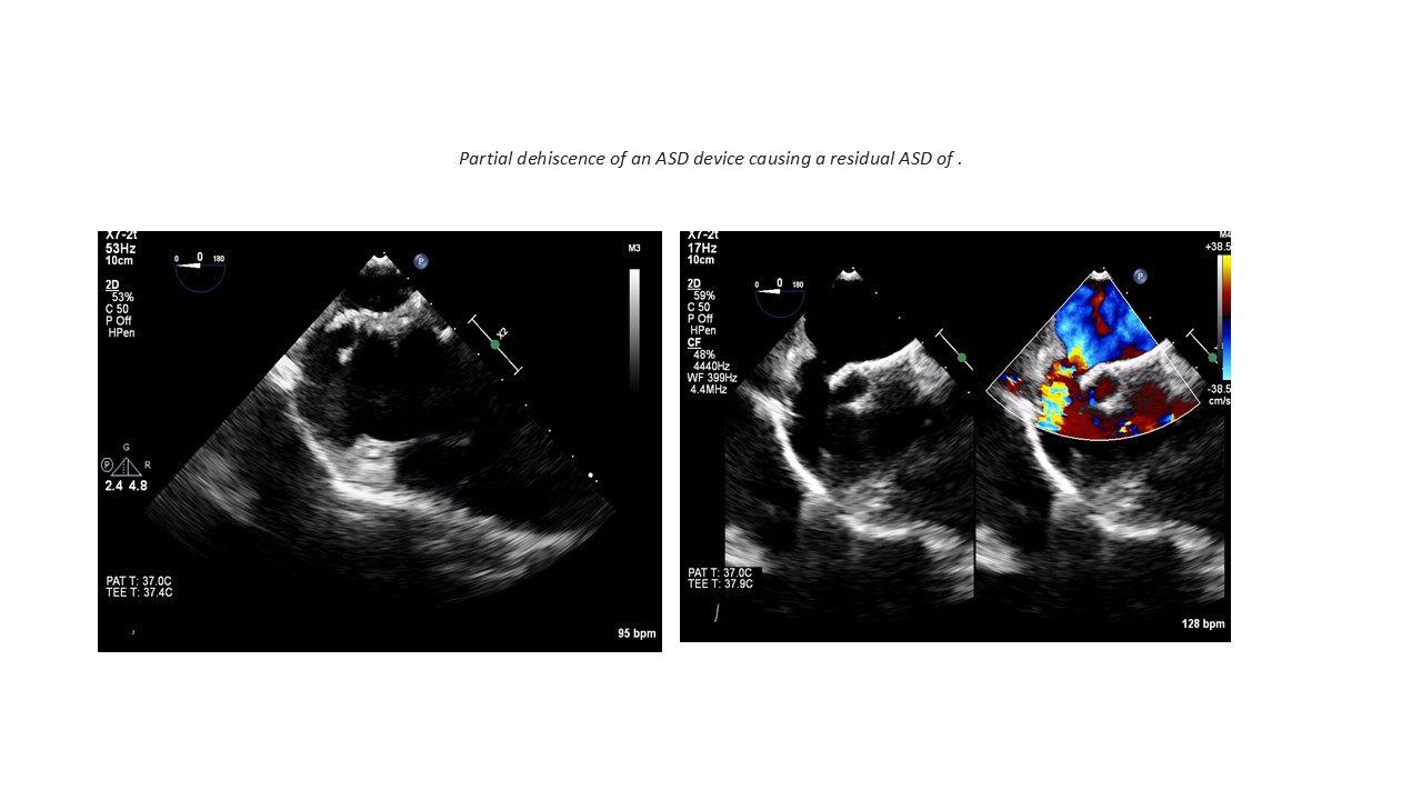
Chest X-ray showed cardiomegaly consistent with an intracardiac shunt; the previous ASD device was visible. ECG demonstrated an incomplete right bundle branch block. Echocardiography revealed the device was oscillating with evidence of a residual left-to-right shunt, suggesting incomplete closure or device-related complication.
Relevant Catheterization Findings
Left and right heart catheterization showed a step-up in oxygen saturation from mixed venous blood to the right atrium, consistent with a residual ASD. Mean pulmonary artery pressure indicated mild pulmonary hypertension. These findings confirmed a hemodynamically significant left-to-right shunt.
Interventional Management
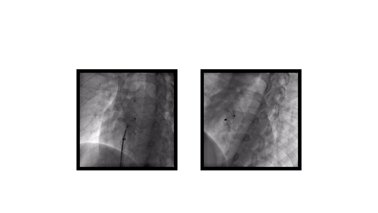
Procedural Step
The patient underwent transcatheter closure under conscious sedation. Femoral venous access was obtained, and a multipurpose catheter was advanced into the right atrium under fluoroscopic and TEE guidance. The residual ASD was crossed with a guidewire, followed by balloon sizing to assess the defect. A second Amplatzer septal occluder device was selected based on defect size and rim assessment. The delivery sheath was positioned across the defect, and the left atrial disc was deployed, followed by the right atrial disc. Proper positioning and device stability were confirmed with TEE, ensuring no impingement on surrounding structures. No significant residual shunt was observed. Once confirmed, the device was released. The final assessment showed successful closure without complications. The patient remained stable throughout the procedure.
 ASD procedure.mp4
ASD Echo.mp4
Case Summary
m a
- Septum condition and anatomy are critical factors in the success of deploying a second device for residual ASD closure.
- Adequate septal tissue, careful imaging assessment, and precise device deployment are essential to ensure a stable and effective closure while minimizing the risk of complications.
- Regular follow-up is necessary to monitor the integrity of the septum and the position of the devices over time.
nd the position of the devices over time.